He aquí unas cuantas micrografías electrónicas que son verdaderamente impresionantes.
Fingerprint texture of cholesteric liquid crystal in a wedge cell
1 February 2012K
en Ishikawa / Tokyo Institute of Technology

A simple snowflake (x4) by Yanping Wang, Beijing Planetarium
1 January 2012
Yanping Wang / Beijing Planetarium

Direct surface view of a charge coupled device sensor
1 March 2012
Kevin Smith / Metprep Ltd

SEM of a section of leaf from a Christmas rose (Helleborus niger)
1 December 2011
Science Photo Library

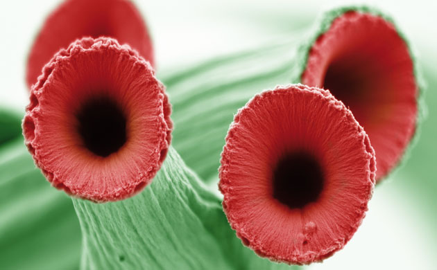
False-coloured SEM of carbon nanotube pillars that look like poppies
1 November 2012
Adrianus / ARIA, California Institute of Technology / Materials Research Society
Confocal micrograph showing bacillus subtilis growth patterns
1 October 2012
F Federici / T Rudge / PJ Spencer / J Haseloff / Wellcome Trust Images

SEM depicting a baby giraffe formed within a jungle of Ni-Al-C dendrites
1 September 2012
Shaahin Amini / Reza Abbaschian / University of California Riverside

SEM of 'microneedles' made from biodegradable polymer
1 August 2012
Peter DeMuth / Wellcome Trust Images

A false-coloured SEM showing caffeine crystals
1 July 2012
Annie Cavanagh / David McCarthy / Wellcome Trust Images

Human dermal fibroblast cultured onto polystyrene surface
1 June 2012
Sergio Bertazzo / Thomas von Erlach / Imperial College London

SEM of a single red blood cell on the tip of a needle
1 November 2011
Science Photo Library

A clutch of unidentified butterfly eggs on a raspberry plant
1 September 2011
www.funnydust.blogspot.com

SEM of the underside of a sticking plaster
31 July 2011
Wellcome Trust Images

Zeiss Apotome microscopy showing murine neurons
1 May 2012
Andrea Caprini and Fabrizio Gelain

HeLa cancer cells using 2-photon fluorescence
1 April 2012
Thomas Deerinck / University of California-San Diego

Todas las imágenes han sido vistas en esta web
Preciosas
ResponderEliminar